In 2026, biological sample preservation is becoming more precise, traceable, and application-specific. Research teams now compare cryopreservation, vitrification, lyophilization, chemical stabilization, and controlled-temperature storage. Each method protects different biological features. Cells may require gradual cooling, while RNA often needs immediate stabilization. Tissue samples demand careful control of ischemic time, moisture, and fixation conditions.
As biobank expert Jan-Eric Litton has stated, “The quality of a biobank depends on the quality of its samples.” This principle remains practical. A sample stored at minus 80 degrees Celsius can still become unreliable after repeated thawing. A liquid nitrogen system can preserve cellular activity, but only with validated monitoring and emergency planning. Small details matter, including tube compatibility, barcode readability, freezer mapping, and temperature records. They are not administrative decoration.
This guide examines the leading biological sample preservation methods expected to shape laboratory practice in 2026. It considers sample type, storage duration, equipment demands, recovery quality, and long-term costs. It also discusses quality control, contamination prevention, and documented chain-of-custody procedures. No method fits every specimen. That limitation deserves attention. Some laboratories still select familiar techniques without sufficient validation. Better decisions begin with pilot testing, reference materials, and documented freeze-thaw studies. Even strong protocols can fail through human error. Reliable preservation therefore combines science, equipment, training, and continuous review.
Preservation protects a sample’s structure, chemistry, and biological signals from collection through analysis. The right method depends on the study’s purpose. DNA, RNA, proteins, living cells, and tissue sections each require different controls. A method that preserves genetic material may damage cell morphology. No single option works for every sample.
Common approaches include controlled freezing, cryopreservation, chemical fixation, dehydration, and validated stabilizing solutions. Temperature control remains essential. Even brief warming can alter enzyme activity or weaken cell recovery. For tissue morphology, fixation may provide clearer microscopic detail. For viable cells, however, fixation is usually unsuitable. Sample volume, container type, storage duration, and transport conditions also influence the choice.
Selection should begin with the final test, not the storage device. Define acceptable quality limits before collection. Record temperature history, collection time, processing delays, and freeze-thaw cycles. These details support traceability and strengthen later interpretation. A small pilot study can reveal hidden problems, such as poor recovery after thawing or uneven tissue penetration. That step is often skipped. It should not be. Preservation plans also need documented procedures, trained personnel, calibrated equipment, and periodic quality checks. In practice, convenience can quietly replace evidence. Reviewing that decision may prevent expensive sample loss.
In 2026, leading preservation methods will depend less on freezer technology and more on disciplined pre-collection planning. A review by Plebani reported that preanalytical errors represented roughly 46–68% of laboratory errors across quality studies. That range is uncomfortable. It shows why collection design deserves equal attention.
Before collection, define the analyte, acceptable delay, temperature window, container, anticoagulant, and minimum volume. ISO 20387:2018 emphasizes traceability, documented competence, and controlled biobanking processes. ISBER Best Practices recommends recording collection time, processing time, deviations, and freeze-thaw history. Label tubes before collection begins. Prepare a chilled rack, calibrated thermometer, absorbent materials, and a backup transport route. Keep the checklist visible.
Handling should match the specimen, not convenience. Whole blood may need gentle inversion, while tissue often requires rapid stabilization and controlled freezing. Serum should clot within a validated interval before separation. Avoid repeated transfers. Record every handoff. One unclear timestamp can weaken an otherwise valuable sample. Short delays matter.
A perfect protocol rarely survives a crowded clinic. Staff may forget one field. Equipment may drift. These weaknesses deserve review, not concealment. Temperature logs, deviation records, and periodic competency checks can reveal where the workflow actually fails. The best preservation decision often begins before the sample exists.
| Sample Type | Recommended Preservation Method | Typical Storage Temperature | Pre-Collection Planning Requirements | Handling Requirements After Collection | Key Quality Risks | Priority |
|---|---|---|---|---|---|---|
| Whole Blood for DNA | Collect in an EDTA tube. For longer storage, separate leukocyte-rich material or use a validated DNA stabilization system. | 2–8°C short term; −20°C to −80°C for extracted DNA | Confirm anticoagulant compatibility, required blood volume, participant identification, and downstream DNA extraction workflow. | Mix gently by inversion. Avoid vigorous shaking and repeated freeze–thaw cycles. Process according to the validated laboratory timeline. | Clotting, hemolysis, leukocyte degradation, incorrect anticoagulant, and repeated temperature excursions. | High |
| Plasma | Collect blood with a validated anticoagulant, centrifuge to separate plasma, aliquot, and freeze promptly. | 2–8°C during short processing; −80°C preferred for long-term storage | Define anticoagulant choice, centrifugation conditions, processing window, aliquot volume, and analyte-specific stability requirements. | Keep samples cool during processing, avoid disturbing the cell layer, use low-binding or validated tubes when required, and minimize headspace where appropriate. | Cellular contamination, delayed processing, platelet activation, evaporation, freeze–thaw damage, and hemolysis. | High |
| Serum | Allow blood to clot under controlled conditions, centrifuge, aliquot, and freeze promptly after separation. | 2–8°C during short processing; −80°C preferred for long-term storage | Specify clotting time, tube type, centrifugation parameters, target analytes, and acceptable hemolysis or lipemia limits. | Do not freeze before clot formation is complete. Separate serum from cells, label aliquots clearly, and avoid repeated thawing. | Incomplete clotting, fibrin, hemolysis, prolonged room-temperature exposure, and analyte degradation. | High |
| Peripheral Blood Mononuclear Cells | Isolate using density-gradient separation, then preserve as a controlled-rate frozen cell suspension with a validated cryoprotective medium. | 2–8°C before processing; vapor-phase liquid nitrogen for long-term storage | Plan same-day processing, cell-count and viability targets, cryoprotectant preparation, controlled-rate freezing, and storage capacity. | Process promptly, maintain aseptic technique, record cell concentration and viability, freeze at a controlled rate, and transfer to appropriate cryogenic storage. | Low viability, delayed processing, osmotic injury, contamination, uncontrolled freezing, and temperature shock. | High |
| Tissue for Histology | Fix promptly in an adequate volume of neutral buffered formalin, followed by validated tissue processing and paraffin embedding. | Room temperature during fixation; embedded blocks at controlled room temperature | Define tissue dimensions, fixation duration, orientation, sampling plan, and whether molecular testing may also be required. | Use a fixative-to-tissue volume ratio of approximately 10:1 where applicable. Avoid excessive thickness and document warm or cold ischemia time. | Autolysis, under-fixation, over-fixation, poor penetration, loss of orientation, and compromised nucleic acid quality. | High |
| Fresh-Frozen Tissue | Snap-freeze rapidly using a validated approach and store in a sealed, labeled cryogenic container. | −80°C for many workflows; vapor-phase liquid nitrogen for extended storage | Predefine tissue sampling, ischemia limits, specimen size, biosafety controls, and whether paired fixed tissue is needed. | Remove excess fluid where appropriate, freeze quickly, prevent thawing during transfer, and record storage location and temperature history. | RNA degradation, freeze–thaw damage, cross-contamination, specimen misidentification, and delayed freezing. | High |
| Saliva for Microbial or DNA Testing | Collect in a sterile container or validated stabilizing collection device according to the intended assay. | 2–8°C if processed promptly; −20°C to −80°C for longer storage, depending on the assay | Control eating, drinking, smoking, oral hygiene, and medication restrictions when required by the study protocol. | Avoid contamination, record collection time, mix only as directed, and follow the validated transport and stabilization limits. | Food or drink contamination, low volume, microbial overgrowth, delayed stabilization, and insufficient participant compliance. | Medium |
| Urine | Collect in a sterile, leak-resistant container. Use preservatives only when validated for the target analyte or microbiology method. | 2–8°C for short-term holding; freezing for longer storage when compatible with the assay | Define collection type, timing, volume, preservative requirements, and whether the specimen is intended for chemistry, nucleic acid, or culture testing. | Mix gently, aliquot when needed, protect light-sensitive analytes, and process or cool promptly to limit bacterial growth and chemical change. | pH changes, bacterial growth, cellular degradation, precipitation, evaporation, and light exposure. | Medium |
| Cerebrospinal Fluid | Use sterile, low-binding containers when appropriate and process immediately for cellular, microbiological, biochemical, or molecular testing. | Room temperature for immediate cellular analysis; 2–8°C or freezing according to assay requirements | Coordinate collection with the testing laboratory, define minimum volume, tube allocation, and urgent transport procedures. | Prevent delays, avoid unnecessary agitation, protect from contamination, and divide into aliquots when multiple tests are required. | Rapid cellular lysis, low-volume loss, contamination, adsorption to surfaces, and inappropriate freezing. | High |
| Microbiological Swabs | Use a sterile swab and transport medium validated for the suspected organism and intended test. | Usually 2–8°C if transport is delayed; follow organism-specific requirements | Choose the correct anatomical site, swab type, transport medium, collection sequence, and timing relative to antimicrobial treatment. | Collect before antimicrobial therapy when clinically appropriate, avoid touching non-target surfaces, maintain the required temperature, and transport promptly. | Inadequate specimen site, desiccation, overgrowth, oxygen exposure, delayed transport, and antimicrobial carryover. | High |
| RNA Samples | Stabilize immediately with a validated RNA preservation reagent or rapidly freeze the specimen. | −80°C for long-term storage; liquid nitrogen may be used for suitable materials | Prepare an RNase-controlled workspace, pre-label tubes, define tissue mass or cell number, and establish a short collection-to-stabilization interval. | Use RNase-free materials, wear clean gloves, keep samples cold, avoid repeated freeze–thaw cycles, and document stabilization time. | RNase contamination, delayed stabilization, thawing, low input quantity, and degradation during extraction. | High |
| Protein or Metabolite Samples | Separate the relevant biological fraction quickly, aliquot, and freeze under conditions validated for the target molecule. | 2–8°C during short processing; −80°C commonly used for long-term storage | Identify stability-sensitive analytes, define light and oxygen controls, select compatible collection tubes, and establish acceptable processing times. | Keep samples cold, minimize exposure to air and light when relevant, avoid repeated thawing, and use consistent aliquot sizes. | Proteolysis, oxidation, adsorption, evaporation, hemolysis, and matrix-related interference. | High |
| Dried Blood Spots | Apply an adequate blood volume to approved collection paper, allow spots to dry completely, and store with desiccant when required. | Dry, dark, controlled room temperature for validated periods; colder storage may be required for sensitive analytes | Define spot size, drying time, humidity limits, target analytes, paper type, and packaging requirements. | Do not stack wet cards, protect from humidity and direct light, use breathable drying conditions, and package only after complete drying. | Incomplete drying, humidity exposure, uneven blood distribution, insufficient volume, and analyte instability. | Medium |
| Cultured Mammalian Cells | Preserve as a viable cell suspension using a validated cryoprotective medium and controlled-rate freezing. | −80°C temporarily; vapor-phase liquid nitrogen for long-term storage | Confirm cell identity, mycoplasma status, passage range, viability target, density, and recovery plan before freezing. | Harvest during the intended growth phase, record viability and cell count, freeze at a controlled rate, and avoid prolonged exposure to cryoprotectant at room temperature. | Contamination, genetic drift, low viability, over-confluence, cryoprotectant toxicity, and poor recovery. | High |
| Fecal Samples | Collect in a sterile, leak-resistant container. Use a validated stabilizer when molecular or microbiome testing requires delayed processing. | 2–8°C for short-term transport; −80°C for long-term storage when compatible with the study design | Define collection amount, diet or medication metadata, anaerobic requirements, stabilizer compatibility, and time-to-freezing limits. | Avoid urine or water contamination, homogenize only when required, aliquot promptly, and limit freeze–thaw cycles. | Microbial composition changes, oxygen exposure, moisture variation, contamination, and delayed freezing. | High |

Note: Storage temperatures and processing windows are general planning ranges, not universal specifications. Final requirements must be confirmed against the intended assay, specimen type, applicable biosafety procedures, validated laboratory protocols, and study-specific quality controls.
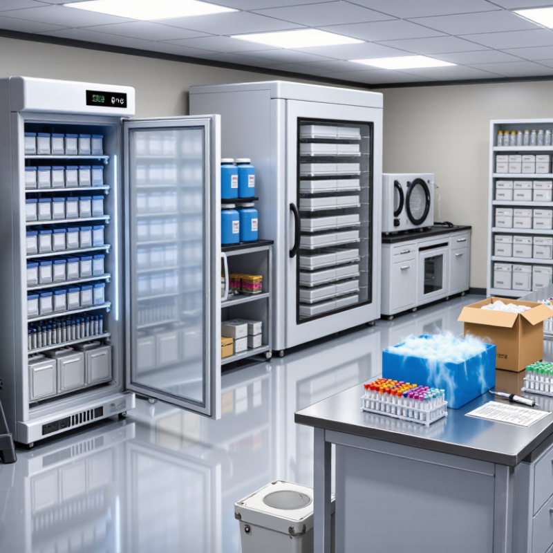
Biological sample preservation should match the intended assay, not merely the specimen. Cells usually need controlled-rate freezing with validated cryoprotectants and rapid transfer to ultra-low temperatures. Vapor-phase cryogenic storage can reduce temperature variation during retrieval. ISBER Best Practices recommend documented temperature monitoring, backup systems, and traceable sample handling.
Tissues require a different decision. Fresh-frozen tissue protects many molecular targets, while formalin-fixed paraffin-embedded tissue supports long-term morphology review. However, fixation time can alter nucleic acids and protein signals.
Biofluids, including plasma, serum, urine, and cerebrospinal fluid, benefit from prompt processing and small aliquots. Repeated freeze-thaw cycles can change concentration measurements. A 2023 national biobank annual report documented specimens from more than 500,000 participants, showing the scale required for consistent storage, labeling, and retrieval. Large collections magnify small procedural errors.
Tips: Define the downstream test before collection. Record collection time, processing delay, temperature history, storage position, and freeze-thaw count. Use reference materials when validating a new protocol. No method is perfect. Even cryogenic storage cannot repair poor pre-analytical handling. My practical bias is to preserve fewer samples properly, rather than store many samples with uncertain histories. Review failure records regularly. That uncomfortable step often reveals hidden weaknesses.
2026 Top Biological Sample Preservation Methods?
Storage Conditions, Monitoring, and Quality Control
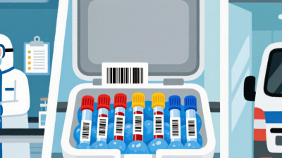
Reliable preservation begins with matching storage conditions to the sample’s biology. Fresh tissues, blood fractions, nucleic acids, and cultured cells need different temperature ranges and handling times. Use validated containers, clear labels, and defined maximum exposure periods. Minimize repeated freeze–thaw cycles by preparing small, documented aliquots. Cold-chain delays can damage samples before they reach long-term storage.
Monitoring must be continuous, not occasional. Record temperature, humidity, equipment status, and door openings with calibrated instruments. Set alerts for gradual drift, not only major failures. A backup power plan and a second storage location reduce avoidable losses. Staff should review alarm responses during training, because an alert is useless if nobody knows who acts.
Quality control should include identity checks, contamination screening, concentration measurements, and viability testing when relevant. Compare results with acceptance limits before releasing samples for research. Keep an auditable record of collection time, processing steps, storage location, and every transfer. No protocol is perfect. Small labeling errors still occur, especially during busy shifts. Periodic audits, independent review, and honest deviation reports help reveal weak points. Some laboratories also retain reference aliquots for later comparison, although this adds space, cost, and another handling risk.
Biological sample preservation is moving beyond standard −80°C freezers. The 2024 Biobanking Market report estimates the sector could grow from about $72.8 billion in 2024 to $95.3 billion by 2029. That expansion reflects rising demand for reproducible tissue, blood, cell, and nucleic-acid storage. Cold chains remain fragile. Power interruptions, door openings, and temperature drift can quietly damage samples before anyone notices.
In 2026, vitrification, controlled-rate freezing, and lower-toxicity cryoprotectant systems will receive greater attention. Vitrification rapidly converts water into a glass-like state, reducing damaging ice crystals in oocytes, embryos, and sensitive cell models. Trehalose-based formulations and dry-state stabilization may also support short-term ambient transport. A 2024 logistics technology report identified real-time temperature monitoring as a major priority across life-science distribution, especially for decentralized clinical studies. Small sensors inside transport containers can record every thermal event, not merely the final temperature.
Artificial intelligence will increasingly flag abnormal freezer patterns and predict maintenance needs. That sounds impressive, but prediction is not preservation. Validation still requires recovery testing, viability measurements, and documented chain-of-custody controls. The 2023 ISBER Best Practices emphasizes standardized procedures, qualified equipment, and traceable records across biobanking workflows. I would not accept a new preservation medium because it performs well in one laboratory. Donor variability, sample volume, repeated thawing, and storage duration can change the result. The uncomfortable gap remains: many emerging methods have promising short-term data, but limited evidence after five or ten years.